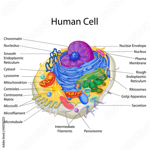
42 diagram of a human cell with labels
Labelled Diagram Of A Human Cell Bone Cell Labeled Diagram Animal Cell ... Labelled Diagram Of A Human Cell Bone Cell Labeled Diagram Animal Cell Free Printable To Label Find this Pin and more on Biology by Guinahi Douhe. Cell Structure Structure And Function Eukaryotic Cell Plasma Membrane Classroom Management Tips Label Image Animal Cell Application Letters Plant Cell More information ... More information cell diagram to label Also, Plant Cell Diagram No Labels Functions - Cell Diagram and also Draw a diagram of human sperm. Label only those parts along with their. [DIAGRAM] Cell Diagram Labeling diagramcloud.blogspot.com labeling labeled teacherspayteachers Chapter 3 Review
Free Cell Diagram Software with Free Templates - EdrawMax - Edrawsoft Before making a cell diagram on EdrawMax, first gather all the necessary supporting facts to draw the diagram. Draw all the cell components roughly into the shape of a cell. The cell wall, cell membrane, cytoplasm, nucleus, and cell organelles are components. Step 2: Template selection. Step 3: Customize the Diagram.
Diagram of a human cell with labels
The Human Skeleton: All You Need to Know - Bodytomy Labeled Skeleton Diagram This skeleton diagram will help explain the different bones of the human body clearly. Cranium The cranium is a skull bone that covers the brain, as seen in the skeleton diagram. The facial bones are not a part of the cranium. The bones that are just above the ear or in front of the ear are known as temporal bones. Stapes Anatomy and Physiology: Parts of a Human Cell - Visible Body Cells can be divided into four groups: somatic, gamete, germ, and stem. Somatic cells are all the cells in the body that aren't sex cells, like blood cells, neurons, and osteocytes. Gametes are sex cells that join together during sexual reproduction. Germ cells produce gametes. Labeled diagram of the human kidney royalty-free images - Shutterstock Labeled diagram of the human kidney royalty-free images 189 labeled diagram of the human kidney stock photos, vectors, and illustrations are available royalty-free. See labeled diagram of the human kidney stock video clips Image type Orientation People Artists Sort by Popular Healthcare and Medical Anatomy Diseases, Viruses, and Disorders kidney
Diagram of a human cell with labels. Labeled Diagrams of the Human Brain You'll Want to Copy Now All the functions are carried out without a single glitch and before you even bat an eyelid. The following are the different regions of the human brain and their functions. Labeled Diagrams of the Human Brain Central Core The central core consists of the thalamus, pons, cerebellum, reticular formation and medulla. Labeled Plant Cell With Diagrams | Science Trends The parts of a plant cell include the cell wall, the cell membrane, the cytoskeleton or cytoplasm, the nucleus, the Golgi body, the mitochondria, the peroxisome's, the vacuoles, ribosomes, and the endoplasmic reticulum. Parts Of A Plant Cell The Cell Wall Let's start from the outside and work our way inwards. Learn the parts of a cell with diagrams and cell quizzes For this exercise we'll start with an image of a cell diagram ready labeled. Study this and make sure that you're clear about which structure is found where. Cell diagram unlabeled It's time to label the cell yourself! As you fill in the cell structure worksheet, remember the functions of each part of the cell that you learned in the video. Draw a labelled diagram of human cheek cells. [3 MARKS] - Byju's Draw a labelled diagram of human cheek cells. [3 MARKS] Solution Squamous epithelium is composed of thin and flat cells, with closely packed nuclei. ∙This type of epithelium is found in the lining of the mouth and nasal cavities, blood vessels, and lymph vessels. Biology Standard VI Suggest Corrections 18 Similar questions Q.
blank cell diagram to label blank cell diagram to label blank cell diagram to label 12 best images of human brain diagram worksheet. The animal cell worksheet. Cycle nitrogen carbon diagram biology biol oakleys understanding blank cell diagram to label Cell Organelles- Definition, Structure, Functions, Diagram A cell wall is multilayered with a middle lamina, a primary cell wall, and a secondary cell wall. The middle lamina contains polysaccharides that provide adhesion and allow binding of the cells to one another. After the middle lamina is the primary cell wall which is composed of cellulose. Label Diagram Human Body Illustrations & Vectors - Dreamstime Download 207 Label Diagram Human Body Stock Illustrations, Vectors & Clipart for FREE or amazingly low rates! New users enjoy 60% OFF. 191,348,773 stock photos online. ... Animal cell structure anatomy infographic diagram. With parts flat vector illustration design for biology science education school book concept microbiology. › articles › viewJCI - Human midbrain dopaminergic neuronal differentiation ... Jun 14, 2022 · Mean gene expression has been scaled between 0 and 1. Horizontal bars denote the number of cells in each cluster. Cell-type labels are used as UMAP clusters in B. The dot color scale represents average expression levels, and dot size represents the fraction of cells in a group.
Blood Cell Diagram Pictures, Images and Stock Photos Blood stem cell is an immature cell that can develop into all types of blood cells, including white blood cells, red blood cells, and platelets. Blood stem cells are found in the peripheral blood and the bone marrow. Also called hematopoietic stem cell. 3d render. Multiple myeloma. plasma cell myeloma. byjus.com › biology › skin-diagramSkin Diagram with Detailed Illustrations and Clear Labels - BYJUS Skin Diagram The largest organ in the human body is the skin, covering a total area of about 1.8 square meters. The skin is tasked with protecting our body from external elements as well as microbes. 03 Label the Cell Diagram | Quizlet Start studying 03 Label the Cell. Learn vocabulary, terms, and more with flashcards, games, and other study tools. ... cell diagram. 18 terms. lugo_janet. Sets found in the same folder. 03 Organelle Functions. 14 terms. muskopf1. 07 Cell Labeling. 11 terms. muskopf1. 03 Cell Transport. 12 terms. Human Cell Organelles Labeling Diagram | Quizlet Human Cell Organelles Labeling STUDY Learn Flashcards Write Spell Test PLAY Match Gravity Created by Mackenna_Rios5 Terms in this set (8) Vesicles Transports molecules between organelles and the cell membrane Ribosome Makes Protein Mitochondria Makes ATP Smooth ER Makes lipids and vesicles Lysosomes
Liver Diagram with Detailed Illustrations and Clear Labels - BYJUS Liver Diagram with Detailed Illustrations and Clear Labels Biology Important Diagrams Liver Diagram Liver Diagram The liver is one of the most important organs in the human body. Anatomically, the liver is a meaty organ that consists of two large sections called the right and the left lobe.
Cells Diagram | Science Illustration Solutions - Edrawsoft Cells Diagram Symbols Edraw software offers you lots of symbols used in cells diagram like cell structure, paramecium, squamous cell, cell division, bacteria, cell membrane, eggs, sperm, zygote, an animal cell, SARS, tobacco mosaic, adenovirus, coliphage, herpesvirus, AIDS, pollen, plant cell model, onion tissue, etc. Cells Diagram Examples
PDF Human Cell Diagram, Parts, Pictures, Structure and Functions Diagram of the human cell illustrating the different parts of the cell. Cell Membrane The cell membraneis the outer coating of the cell and contains the cytoplasm, substances within it and the organelle. It is a double-layered membrane composed of proteins and lipids.
blank cell diagram to label cell labeling ks2 Animal Cell Diagram - Unlabeled - Tim's Printables unlabeled labeled Blank cell diagrams. 31 label a cell diagram. Labeling lymph node lymphatic system structures vessel efferent section appropriate chapter immunity labels correctly match terms below each card through

Questions And Answers On Labeled/Unlebled Diagrams Of A Human Cell / Questions And Answers On ...
Human Cells Printables and Diagrams - The Successful Homeschool These cells include: leukocytes, haematids, thrombocytes, ovum, sperm, sarcomeres, enterocytes, neurons, osteocytes, hepatocytes. They will learn the parts of a cell thanks to a labeled diagram. They will get to see what blood looks like under a microscope without needing to own a microscope. They get to color a cell and then label the parts.

Animal Cells Diagram with Labels Awesome Animal Cell Diagrams Labeled | Animal cell project ...
Animal Cells: Labelled Diagram, Definitions, and Structure - Research Tweet Animal Cells Organelles and Functions. A double layer that supports and protects the cell. Allows materials in and out. The control center of the cell. Nucleus contains majority of cell's the DNA. Popularly known as the "Powerhouse". Breaks down food to produce energy in the form of ATP.
Structure of Cell: Definition, Types, Diagram, Functions - Embibe Structure of Cell: Cell is the basic functional unit that makes up all living organisms.All organisms, including ourselves, start life as a single cell called the egg. Cells are small microscopic units that perform all essential functions of life and are capable of independent existence.
Anatomy (Human Body) Labeling - Exploring Nature Arteries of the Lower Limb (Pelvis, Leg and Foot) Labeling. Arteries of the Upper Limb (Shoulder, Arm, Hand) Labeling. Blood Vessel Anatomy Labeling
Cell: Structure and Functions (With Diagram) - Biology Discussion Eukaryotic Cells: 1. Eukaryotes are sophisticated cells with a well defined nucleus and cell organelles. 2. The cells are comparatively larger in size (10-100 μm). 3. Unicellular to multicellular in nature and evolved ~1 billion years ago. 4. The cell membrane is semipermeable and flexible. 5. These cells reproduce both asexually and sexually.
Labeled diagram of the human kidney royalty-free images - Shutterstock Labeled diagram of the human kidney royalty-free images 189 labeled diagram of the human kidney stock photos, vectors, and illustrations are available royalty-free. See labeled diagram of the human kidney stock video clips Image type Orientation People Artists Sort by Popular Healthcare and Medical Anatomy Diseases, Viruses, and Disorders kidney
Anatomy and Physiology: Parts of a Human Cell - Visible Body Cells can be divided into four groups: somatic, gamete, germ, and stem. Somatic cells are all the cells in the body that aren't sex cells, like blood cells, neurons, and osteocytes. Gametes are sex cells that join together during sexual reproduction. Germ cells produce gametes.
The Human Skeleton: All You Need to Know - Bodytomy Labeled Skeleton Diagram This skeleton diagram will help explain the different bones of the human body clearly. Cranium The cranium is a skull bone that covers the brain, as seen in the skeleton diagram. The facial bones are not a part of the cranium. The bones that are just above the ear or in front of the ear are known as temporal bones. Stapes


Post a Comment for "42 diagram of a human cell with labels"